
Retinal changes linked to later atrial fibrillation diagnosis
Eye imaging could inform earlier risk assessment, but clinical usefulness remains unproven
Changes in retinal structure were associated with atrial fibrillation in a study reported by the University of Birmingham, led with UCL and Moorfields Eye Hospital. Published in PLOS Digital Health, the research analysed eye images using data from more than 90,000 people.
Thinning in specific retinal layers was also observed in people who later developed atrial fibrillation, on average four years before hospital diagnosis. The association remained significant after adjustment for factors including age, hypertension and diabetes. These findings do not establish causation or demonstrate that eye-based screening improves health outcomes.
Why this matters for policymakers
- Validation Independent testing should establish predictive accuracy before NHS adoption.
- Clinical benefit Evaluation should test whether earlier identification improves outcomes.
- Service planning Any screening proposal needs assessment of referral demand, costs and equitable access.
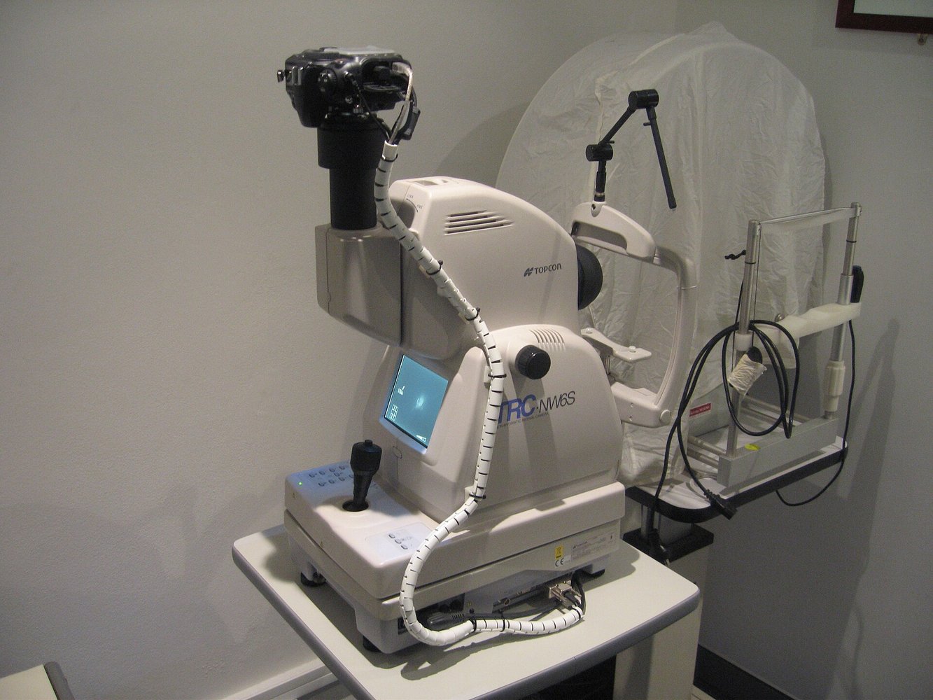
Image: Illustrative photograph of a non-mydriatic retinal camera, not equipment identified as used in this study. Jason Ruck (User:Jason7825), CC BY-SA 3.0. Resized and converted to JPEG for display.